X-ray Bone
A bone X-ray uses a very small dose of ionizing radiation to produce pictures of any bone in the body. It is commonly used to diagnose fractured bones or joint dislocation. Bone X-rays are the fastest and easiest way for your medical provider to view and assess bone fractures, injuries and joint abnormalities.
A bone X-ray makes images of any bone in the body, including the hand, wrist, arm, elbow, shoulder, spine, pelvis, hip, thigh, knee, leg (shin), ankle or foot.
What to Bring
- Your medical provider order (Your medical provider may have already sent this to us.)
- Personal identification
- Insurance card(s)
How to Prepare for an X-ray of Your Bone
- A bone X-ray requires no special preparation.
- You will be asked to remove some of your clothing and to wear a gown during the exam.
- You may also be asked to remove jewelry and any metal objects or clothing that might interfere with the X-ray images. We encourage you to leave valuable jewelry at home.
- If you are pregnant or think you might be, please inform the imaging technologist.
- Many imaging tests are not performed during pregnancy so as not to expose the fetus to radiation. If an X-ray is necessary, precautions (shielding to the abdomen) will be taken to minimize radiation exposure.
- If you have an on-body device (insulin pumps, insulin regulators, Neulasta, other chemo pumps, etc.), you must inform your X-ray technologist. These devices cannot be present in the X-ray room during your exam.
What to Expect
- The X-ray technologist will position you on the X-ray table and place the X-ray imaging plate under the table or directly under the body part in the area of the body being imaged. When necessary, sandbags, sponges or other positioning devices will be used to help you maintain the proper position. A lead apron may be placed over your pelvic area or breasts when feasible to protect from radiation.
- You must hold very still and may be asked to hold your breath for a few seconds while the X-ray picture is taken to reduce the possibility of a blurred image.
- You may be repositioned for another view, and the process is repeated. Two or more images (from different angles) will typically be taken.
- A bone X-ray examination is usually completed within 10-20 minutes, depending on the exam being performed.
Key Points to Know During and After the Exam
- A bone X-ray examination is a painless procedure.
- You may experience discomfort from the cool temperature in the examination room or the hardness of the X-ray table. You may find that the positions you need to hold are uncomfortable, especially if you have an injury.
-
The X-ray technologist will assist you in finding the most comfortable
position possible to ensure diagnostic image quality.
TO SCHEDULE AN APPOINTMENT
Call North Oaks Scheduling between 7 am and 5:30 pm.
Hammond: (985) 230-7777
Livingston: (225) 686-4899If you are a provider and need to send an order, please send it via fax to (985) 230-6781.
-
 North Oaks – Livingston Parish Medical Complex (Diagnostic Services) Livingston Parish 17199 Spring Ranch Road
Livingston, LA 70754
(844) 277-8669 More Information -
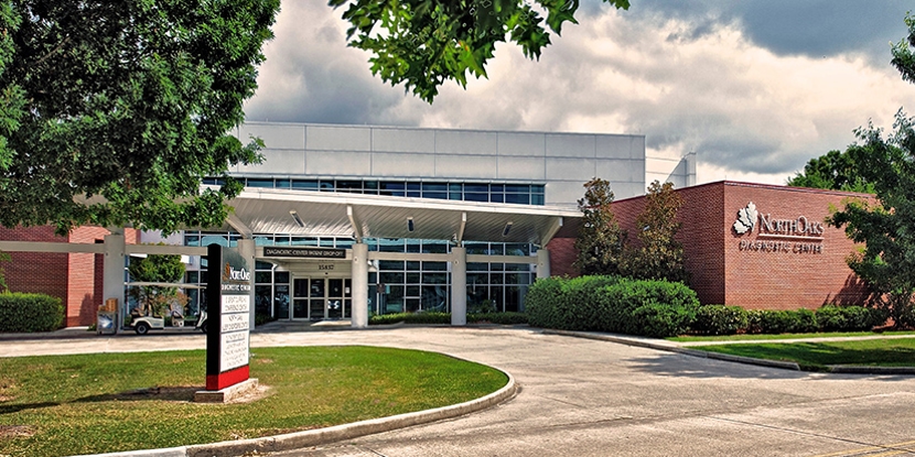
North Oaks Diagnostic Center Screening & Tests 15837 Paul Vega, MD, Drive , Building 1
Hammond, LA 70403
(985) 230-7777 More Information -
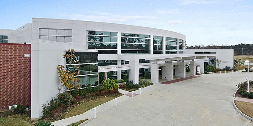
North Oaks Medical Center North Oaks Medical Center 15790 Paul Vega, MD, Drive
Hammond, LA 70403
(985) 345-2700 More Information