Screening Mammogram
Screening mammography is a specific type of breast imaging that uses low-dose X-rays to detect cancer early – before women experience symptoms – when it is most treatable. Mammography plays a central part in the early detection of breast cancers because it can show changes in the breast up to two years before you or your medical provider can feel them. The American Medical Association (AMA) and the American College of Radiology (ACR) recommend annual mammograms for women over 40. The National Cancer Institute (NCI) adds that women who have a personal or family history of breast cancer should talk to their medical provider about when they should begin screening. Screening mammograms are ordered for women with no concerning symptoms such as no lumps, nipple discharge or skin changes.
What to Bring
- Your medical provider order (if you were given an order from your medical provider)
- Personal ID
- Insurance card(s)
How to Prepare
- If you have tender breasts the week before your period, it is best to schedule your mammogram one week following your menstrual cycle.
- If possible, please inform the scheduling department of the location of your prior mammogram so we can request your previous images and have them available for the radiologist to compare at the time of your current mammogram.
- Wear a two-piece outfit.
- Do not use deodorant, talcum powder or lotion under your arms or near the breasts. These products may show up as an artifact in the image.
- Please leave valuable jewelry at home.
- If you are pregnant or think you might be pregnant, please inform the imaging technologist.
- If you wear an on-body device (insulin pump, insulin regulators, Neulasta, other chemo/insulin devices, etc.), you must inform your X-ray technologist. These devices cannot be in the X-ray room during the exam.
- If you have a bruise or a rash on the breast area on the day of your appointment, you may need to reschedule until the area is healed.
What to Expect
- A specially qualified radiological technologist will bring you into a dressing room to verify your information and go over your prior breast history.
- You will be asked to remove your clothing above the waist, and you will be given a cloth gown for the test.
- Once you are in the exam room, the technologist will ask to look at your breast to mark any raised moles, note any scars and check for bruising and/or rashes.
- During your mammography exam, your breast will be placed on a special platform and gradually compressed. The technologist will do their best to maintain your comfort.
-
Breast compression is necessary to:
- Even out the breast thickness so that all tissue can be visualized
- Spread out the tissue so that small abnormalities are less likely to be hidden by overlying breast tissue
- Allow the use of a lower X-ray dose since a thinner amount of breast tissue is being imaged
- Hold the breast still to minimize blurring of the imaged caused by motion
- Reduce X-ray scatter to increase sharpness of the picture
- You will be asked to change positions between images. The routine views are a top-to-bottom view and an angled side view. During positioning, you may be asked to lift your arm or use your hand to hold your other breast out of the way.
- During the exposure you must hold very still and may be asked to hold your breath for a few seconds while the X-ray image is taken.
- You will need to allow 30 minutes from the time you check in to the completion of the test.
- Results are usually available to your ordering medical provider in 24 hours.
- Results will also be mailed to you by the facility.
- Please note that follow-up examinations may be necessary if your screening mammogram shows a potential area of concern. Your radiologist may recommend further diagnostic tests that include additional mammography views and/or ultrasound.
Schedule your 3D mammogram today.
Call North Oaks Patient Scheduling between
7 am and 5 pm, Monday through Friday.
Hammond: (985) 230-7777
Livingston: (225) 686-4899
If you are a medical provider and need to send an order, please fax (985) 230-6781.
-
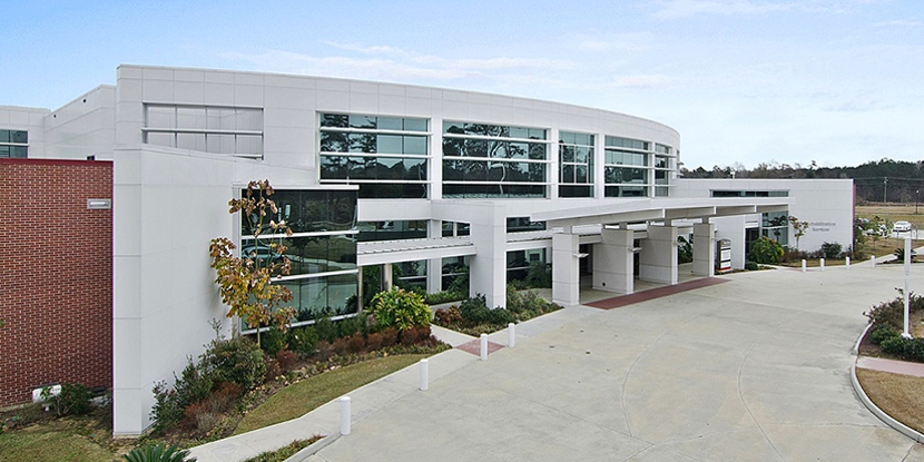
 North Oaks – Livingston Parish Medical Complex (Diagnostic Services) Livingston Parish 17199 Spring Ranch Road
Livingston, LA 70754
(844) 277-8669 More Information -
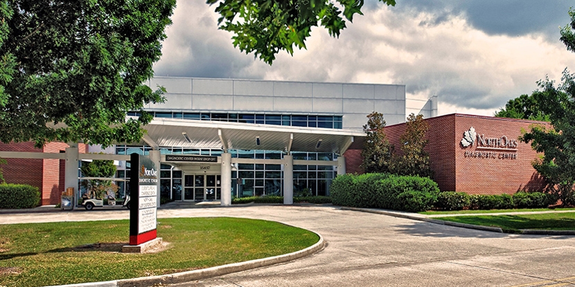
North Oaks Diagnostic Center Screening & Tests 15837 Paul Vega, MD, Drive , Building 1
Hammond, LA 70403
(985) 230-7777 More Information