Lower Extremity Venous Ultrasound
A lower extremity venous ultrasound is a vascular ultrasound that uses ultrasound technology to create images of your blood vessels, such as your veins. This type of ultrasound is primarily used to diagnose deep vein thrombosis (DVT). One or both of your legs may be imaged by ultrasound; it depends on what your medical provider has ordered.
What to Bring
- Your medical provider order (Your medical provider may have already sent this to us.)
- Personal ID
- Insurance card(s)
How to Prepare
- You do not have to do anything special to prepare for a lower extremity venous ultrasound.
- If you have a history of blood clots, you should inform the ultrasound technologist.
- No risks are associated with a lower extremity venous ultrasound. Unlike X-rays, radiation is not involved with this test.
What to Expect
- If you’re wearing slacks, you might be asked to remove them and change into a gown during your exam.
- The room is usually dark so the images can be seen clearly on the computer screen.
- During the exam you will be asked to lie on a table.
- An ultrasound technologist will spread a clear, warm gel on your leg starting at the groin continuing through to your calf. This gel is required to help with the transmission of sound waves through a small wand (transducer). The transducer emits high-frequency sound waves, and a computer measures how the sound waves bounce back from inside the body. The computer changes those sound waves into images to be analyzed.
- The ultrasound technologist will need to apply pressure throughout your leg to ensure there is no evidence of a blood clot.
- Please allow 45 minutes from the time you check in to the completion of the test.
Getting the Results
A radiologist (a physician who is specially trained in reading and interpreting diagnostic and ultrasound images) will interpret the ultrasound results and send the information to your medical provider.
Schedule an Appointment
Call North Oaks Scheduling between 7 am and 5:30 pm.
Hammond: (985) 230-7777
Livingston: (225) 686-4899
If you are a medical provider and need to send an order, please fax it to (985) 230-6781.
-
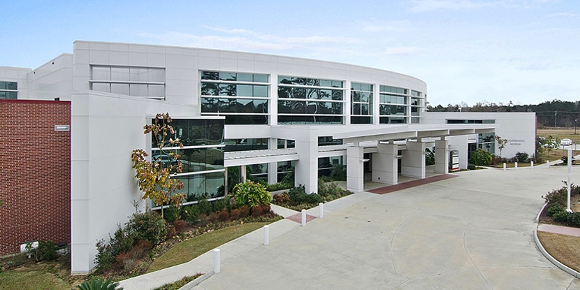
 North Oaks – Livingston Parish Medical Complex (Diagnostic Services) Livingston Parish 17199 Spring Ranch Road
Livingston, LA 70754
(844) 277-8669 More Information -
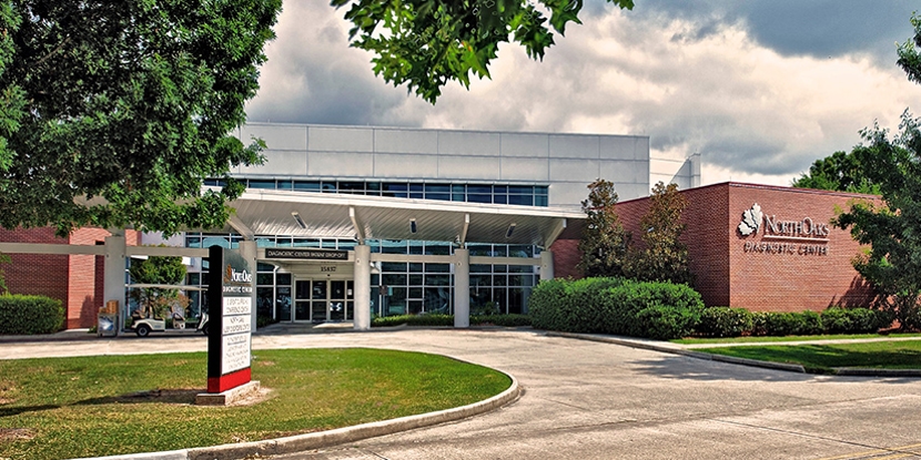
North Oaks Diagnostic Center Screening & Tests 15837 Paul Vega, MD, Drive , Building 1
Hammond, LA 70403
(985) 230-7777 More Information -
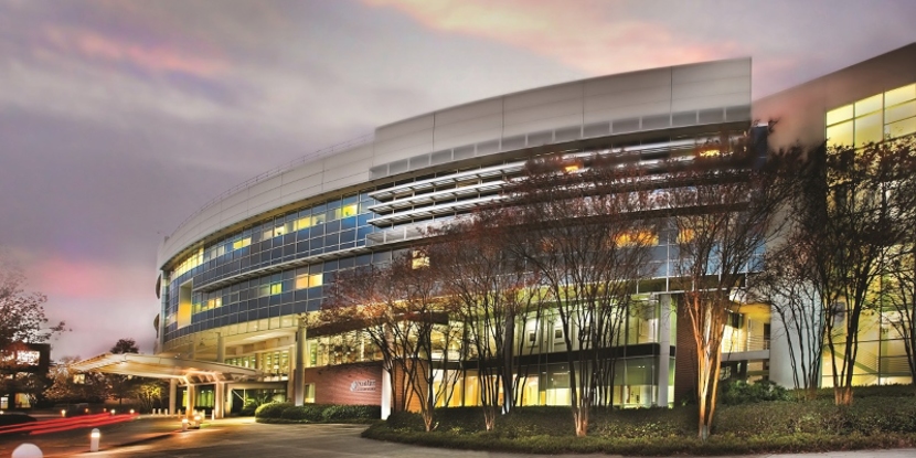
North Oaks Medical Center North Oaks Medical Center 15790 Paul Vega, MD, Drive
Hammond, LA 70403
(985) 345-2700 More Information